baseline vaginal ultrasound
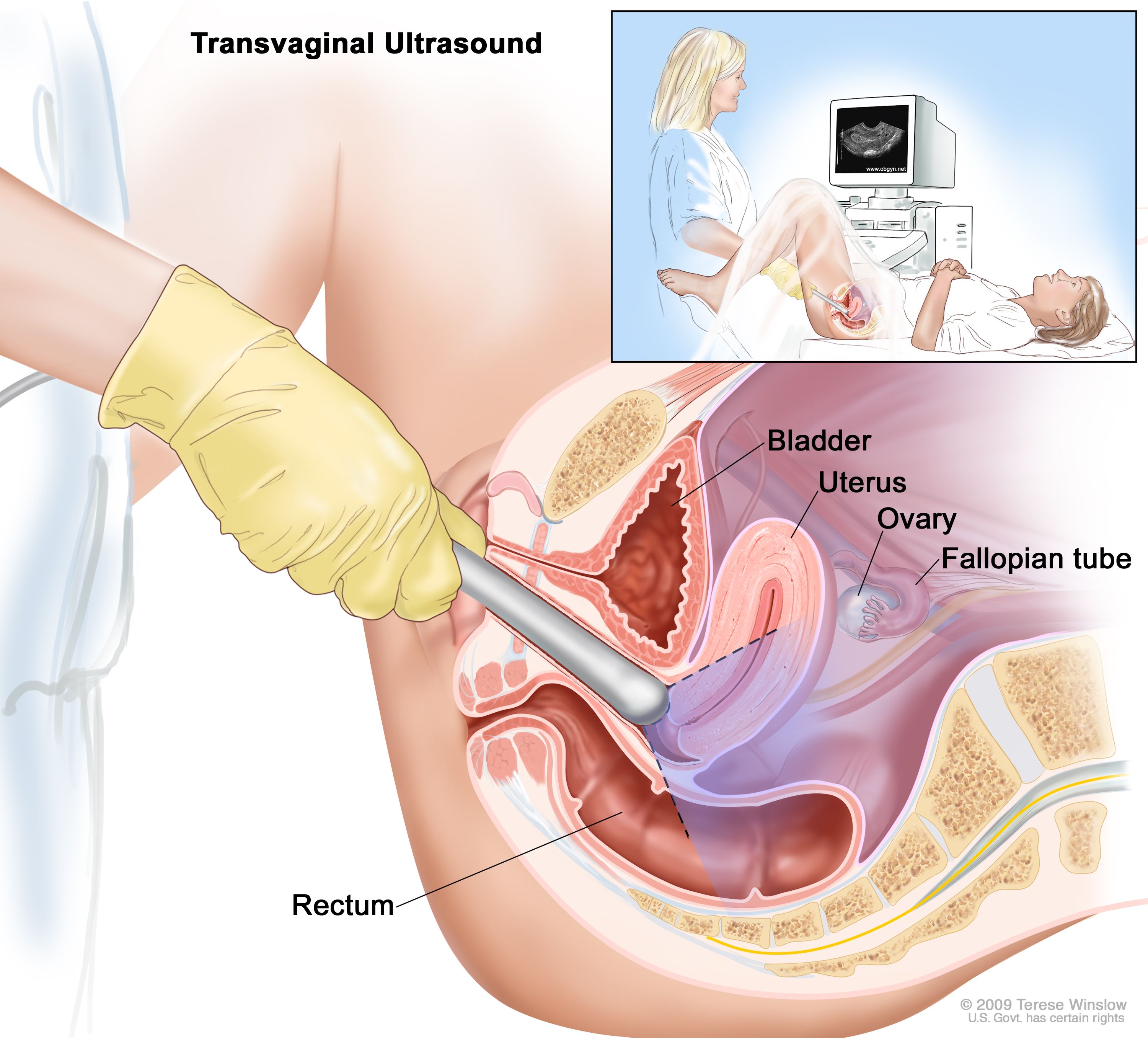
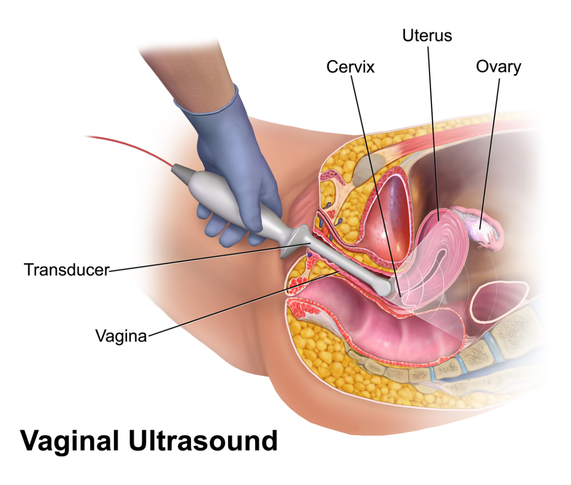
A transvaginal ultrasound can serve many purposes so the goal of yours depends on the type of ultrasound our Las Vegas fertility doctors prescribe. A transvaginal ultrasound gives your fertility care team a look at your reproductive organs including your uterus and ovaries.

Advanced Pelvic Ultrasound In House At Veritas Fertility Surgery
The baseline ultrasound for an intrauterine insemination IUI is the same as a baseline scan for an IVF cycle.

. The posterior vaginal wall is in close proximity to the anterior. Transvaginal ultrasound images showing the pelvis and ovary structures with far greater clarity. Four surgeons performed operations on 85 patients.
Prospective validation by IOTA group. Before initiating tamoxifen baseline vaginal ultrasound sonohysterography or office hysteroscopy to exclude the presence of endometrial polyps appears appropriate. Imaging tests can identify abnormalities and help doctors diagnose conditions.
Basic Tests for Female Patients. The ultrasound is still done on cycle day 2-3 of your menstrual cycle and will help determine if you can start your IUI cycle that day. The general pelvic ultrasound fertility scan generates an image of a number of different important parts of your reproductive tract.
Infertility is defined as the inability to conceive after one year of unprotected sexual intercourse in women under 35 and 6 months of unprotected intercourse in women older than 35. You can do these tests on the same day as your baseline ultrasound. The Basal Antral Follicle Count test is a transvaginal ultrasound study that measures a womans ovarian reserve or her remaining egg supply.
In pre-conception imaging 3D ultrasound is particularly useful for identifying abnormalities in the uterus it helps in the evaluation of the uterine cavity for the presence of endometrial polyps benign growths of the lining of the uterus uterine fibroids benign tumours of the muscle of the uterus or other conditions such as endometriosis. A requisition is needed if you are doing this ultrasound elsewhere. A baseline ultrasound may also be performed prior to a frozen embryo transfer.
Simple ultrasound rules to distinguish between benign and malignant adnexal masses before surgery. When our doctors observe the results of the baseline ultrasound they will be. When a girl reaches puberty.
If an appointment is not available wait for your next cycle. If you are or have been on Tamoxifen Armidex Anastrozole or Femara it is important that you get a baseline ultrasound of your uterus and then follow up with a second ultrasound two years later. A transvaginal ultrasound also called an endovaginal ultrasound is a.
During infertility testing ultrasound scans can provide information on the ovaries endometrial lining and uterus. Infertility affects about 155 of women1 The etiologies of infertility include tubal factor 14 defects in ovulation 21 and male factor 26 and are often unexplained by traditional. Unlike IVF an IUI cycle typically use oral ovulation induction medications like Clomid or.
The ultrasound may take 1530 minutes. The ultrasound is trans-vaginal a wand-like transducer is inserted vaginally to look at your ovaries uterus and. A transvaginal ultrasound showing the heartbeat of a fetus early in a pregnancy.
Call 514 843-1650 on day 1 of your cycle to request an appointment. Transvaginal sonography TVS is a recent addition to the diagnostic techniques available for the evaluation of the female pelvis. Starting a Treatment Cycle.
The lines at the bottom are called M Mode which is how imaging specialists detect and document heart motion. Our experience in over 200 cases of postmenopausal women is the subject of this synoptic review. The baseline ultrasound should be done on day two or day three of your period.
Baseline vaginal ultrasound at the start of the cycle to provide information on uterine and ovarian morphology and to delineate pelvic structures. Your doctor will insert a speculum into your. Baseline creatinine and renal ultrasound findings.
Using this technique in 60 women we were able to detect endometrial changes such as endometrial carcinoma or. These include the uterus also known as the womb the lining of the womb the ovaries and general. The vagina is limited on its outer surface by the hyperechoic visceral facia.
Hysterosonography is performed very much like a gynecologic exam. Timmerman D Ameye L Fischerova D et al. 6th floor of Block C Room C 064351.
On day one of your menstrual period first day of flow not spotting please call CRM to schedule a baseline ultrasound. The colored areas show blood flow. The ovarian reserve reflects her fertility potential.
A transvaginal ultrasound is a safe scan with no known risks. For ultrasound TVS evaluation it is important that the probe once introduced into the vagina is gradually moved upwards while simultaneously evaluating the anterior and posterior vaginal walls. A baseline ultrasound allows us to evaluate the ovaries and pelvic organs to determine if it is an appropriate time to begin ovarian stimulation which is the first phase of IVF treatment.
During fertility treatment ultrasound is used to monitor follicle. This kind of ultrasound provides a clear evaluation of the uterus and ovaries allowing for the diagnosis of uterine masses or ovarian cysts. Hysterosonography also called sonohysterography uses sound waves to produce pictures of the inside of a womans uterus and help diagnose many problems including unexplained vaginal bleeding infertility and repeated miscarriages.
Baseline Pelvic Ultrasound A baseline pelvic ultrasound is an internal vaginal ultrasound which is recommended for most patients as a part of their initial evaluation before starting treatments. People might experience some light discomfort during it but this should go away afterward. Specialized ultrasounds can be used to evaluate ovarian reserves the uterine shape in more detail and whether the fallopian tubes are open or blocked.
The mean creatinine in patients with vesicovaginal fistulas VVF was 060 ngml versus patients with uretero-vaginal fistulas UVF 079 ngml P 0012. Monitoring was started on the 8th day of the. Unlike men who produce sperm on an ongoing basis females are born with a lifetime supply of eggs in their ovaries.
When you are a new patient we often start with a transvaginal.
Ultrasound And Infertility Radiology Key

Definition Of Transvaginal Ultrasound Nci Dictionary Of Cancer Terms Nci

Evaluation Of Quality Of Renal Tract Ultrasound Scans And Reports Performed In Children With First Urinary Tract Infection Journal Of Medical Imaging And Radiation Sciences

Advanced Pelvic Ultrasound In House At Veritas Fertility Surgery

Transverse Ts And Longitudinal Ls Transvaginal Ultrasound Images Of Download Scientific Diagram

Transvaginal Ultrasound Images Showing Typical Features Of A Right Download Scientific Diagram

Sonographic Characterization And Surveillance Of Paravaginal Smooth Muscle Tumor Of Uncertain Malignant Potential Zamora Journal Of Clinical Ultrasound Wiley Online Library

Transvaginal Ultrasound Scan From A Patient With Spontaneous 46 Xx Download Scientific Diagram

Laparoscope And Transvaginal Ultrasound Images During Ablation Download Scientific Diagram

Diagnostics Free Full Text Ultrasound Measurement Of Tumor Free Distance From The Serosal Surface As The Alternative To Measuring The Depth Of Myometrial Invasion In Predicting Lymph Node Metastases In Endometrial Cancer
Gynaecology Ultrasound Women S Ultrasound Melbourne

A Phase Iii Randomised Trial Of Trans Abdominal Ultrasound In Improving Application Quality And Dosimetry Of Intra Cavitary Brachytherapy In Locally Advanced Cervical Cancer Gynecologic Oncology
Gynaecology Ultrasound Women S Ultrasound Melbourne


Comments
Post a Comment